Unlocking New Insights into Cancer Metastasis and Treatment Resistance through Circulating Tumor Cell (CTC) Analysis
Circulating Tumor Cells (CTCs) have emerged as an important research target for understanding two of the greatest challenges in oncology: cancer metastasis and treatment resistance.
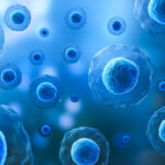
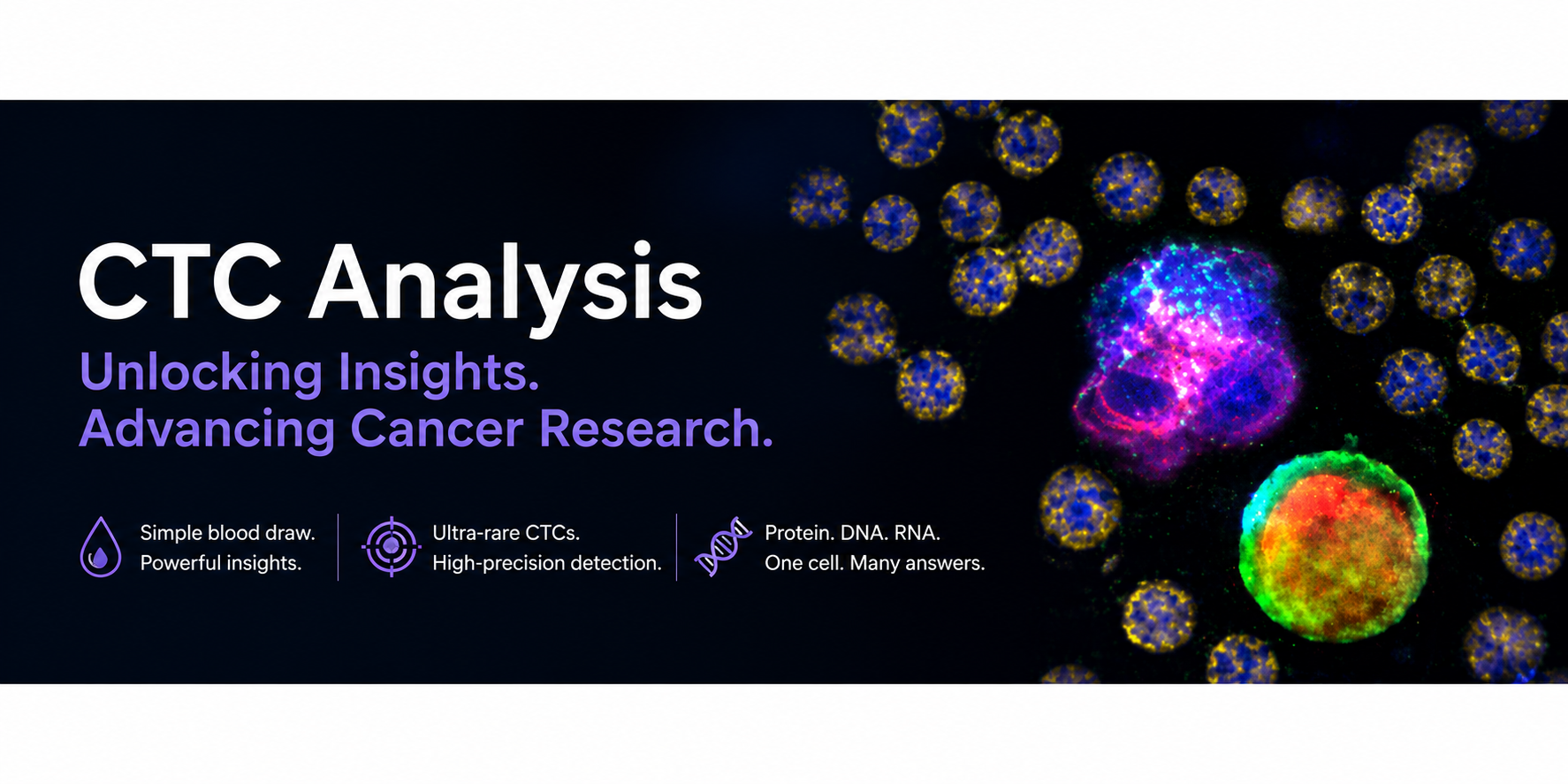
CTCs are tumor cells that have detached from primary or metastatic tumors and entered the bloodstream. Because they can be collected through a simple blood draw, they provide a minimally invasive alternative to tissue biopsy and enable longitudinal monitoring of disease progression and treatment response.
Intact Tumor Cells Obtained from a Blood Sample
Unlike cell-free biomarkers, CTCs remain intact after collection. Their preservation allows researchers to investigate a wide range of biological characteristics, including:
- Mechanisms of cancer progression and metastasis
- Biomarker expression in tumor cells
- Monitoring therapeutic response following treatment
- Evaluation of drug targets and downstream signaling pathways
- Single-cell molecular analysis
Because cellular morphology is preserved, CTCs enable researchers to correlate protein expression with high-resolution imaging, providing valuable phenotypic information in addition to molecular analysis.
Advantages of CTC Analysis Compared with cfDNA
Liquid biopsy commonly includes analysis of circulating cell-free DNA (cfDNA), which is highly useful for detecting tumor-associated genetic alterations. In contrast, CTCs provide access to intact cells, allowing analysis of DNA, RNA, and proteins simultaneously. Interest in protein expression analysis using CTCs has recently increased, particularly in drug discovery and pharmaceutical development. Longitudinal monitoring of therapeutic targets and downstream signaling proteins can help researchers understand target engagement and evaluate whether a drug is performing as intended.
Potential as an Early Indicator of Treatment Response
CTCs are also being investigated as surrogate endpoints in clinical trials. According to RareCyte, changes in CTC number and biomarker expression can sometimes be detected within only a few days after treatment initiation—considerably earlier than changes observed by conventional tumor imaging. Early assessment of therapeutic response may support decision-making in clinical development, including confirmation of mechanism of action, dose optimization, and evaluation of treatment efficacy.
Circulating Tumor Cells: Methods and Protocols
RareCyte’s comprehensive CTC workflow is featured in the Springer Nature publication
Circulating Tumor Cells: Methods and Protocols.
This reference book presents practical laboratory methods for CTC isolation, detection, imaging, biomarker characterization, and single-cell molecular analysis, making it a valuable resource for researchers working in liquid biopsy and cancer biology.

Springer Nature
Comprehensive CTC Analysis with the AccuCyte–CyteFinder Workflow
In Chapter 12 of the book, RareCyte’s scientists describe the comprehensive AccuCyte–CyteFinder workflow for image-based CTC analysis. The workflow integrates every step from blood collection to downstream molecular analysis.
- Blood sample collection
- Sample stabilization and preparation
- Automated staining
- High-resolution imaging
- CTC detection and verification
- Biomarker characterization
- Single-cell isolation
- Downstream DNA and RNA analysis
By combining cellular morphology, high-resolution imaging, biomarker expression, and single-cell isolation, researchers can perform integrated phenotypic and molecular analyses of individual CTCs.
Applications from Early Pharmacodynamic Studies to Target Quantification
According to RareCyte, the AccuCyte–CyteFinder platform is currently being used in clinical trials around the world. Applications range from early pharmacodynamic studies to quantification of therapeutic targets and downstream effector proteins, as well as longitudinal monitoring of biomarker changes during treatment. These capabilities provide researchers with valuable insights into tumor biology, therapeutic response, and mechanisms of drug resistance.
Image-Based CTC Analysis Presented in Chapter 12
Detailed protocols for image-based CTC analysis using the AccuCyte–CyteFinder platform are presented in Chapter 12 of
Circulating Tumor Cells: Methods and Protocols.
The chapter covers the complete workflow, from blood collection and sample stabilization through automated staining, high-resolution imaging, biomarker characterization, single-cell isolation, and downstream molecular analysis.
View the Book on Springer Nature
This article has been prepared for Pharma Nexus based on information published by RareCyte. The analytical performance and potential clinical applications described may vary depending on study design, sample quality, and research objectives.