Beyond the Amino Acid Sequence — Lipid Modifications
How Small Lipids Determine Protein Function
What Light Sensing and Ras Have in Common (Part 6)
In the previous article, we explored how glycans attached to proteins influence encounters between influenza viruses and cells. This time, let us turn our attention to lipids. When we hear the word “lipids,” we may think of energy sources or the building blocks of cell membranes. But lipids can also attach to proteins and change where those proteins work.
What Changes When a Lipid Is Attached to a Protein?
Phospholipids in cell membranes have a water-loving part and a water-repelling part. In water, the water-repelling parts gather on the inside to form a bilayer. This is the familiar lipid bilayer that separates the inside of a cell from the outside.
Now imagine attaching a small, water-repelling lipid chain to a protein dissolved in water. If the chain inserts into the interior of a membrane, it could tether the protein to the membrane, much like an anchor holding a boat in place. This small attachment also plays a part in our ability to see.
An Unexpected Attachment to a Protein That Relays Light Signals
Rod photoreceptor cells in the retina detect faint light in dim surroundings. Rhodopsin is the molecule that absorbs light in these cells. In both humans and cattle, rhodopsin consists of a protein of 348 amino acids bound to 11-cis-retinal, a lipid derived from vitamin A. When retinal absorbs light, one of its double bonds changes from the cis to the trans configuration, producing the all-trans form. This small change in shape triggers a change in the protein as a whole, activating rhodopsin so that it can relay a signal to the inside of the cell.
Transducin receives and amplifies this signal. It consists of three components, or subunits: α, β, and γ. When GDP (guanosine diphosphate) bound to the α subunit is exchanged for GTP (guanosine triphosphate), the α subunit becomes ready to pass the signal to the next molecule. A lipid attached to the small γ subunit plays an important role in this process.
In 1990, Fukada and colleagues reported that a lipid called a farnesyl group was attached to the end of the γ subunit of transducin isolated from bovine retinas (1). The βγ complex carrying this lipid promoted activated rhodopsin-dependent GTP binding to the α subunit much more effectively than did the complex lacking it.
Farnesyl groups had previously been found in yeast mating pheromones, which are peptides. The same attachment was present on molecules that help yeast find a mating partner and on a protein that relays light signals in animal eyes. In the molecular world, shared mechanisms can turn up in unexpected places.
A Small “Anchor” Determines Where a Protein Works
The identity of this attachment was revealed by structural analysis using mass spectrometry. Why, then, had the lipid modification escaped detection? At the time, the amino acid sequence of the γ subunit had already been reported by Ovchinnikov, a prominent biochemist in the former Soviet Union, and his colleagues. Perhaps it was hard to imagine that a protein whose sequence was already known might still carry an unidentified attachment. Also, analyzing the C-terminus and identifying cysteine (Cys) residues were difficult with the protein chemistry methods available at the time. I believe these difficulties also contributed to the modification going undetected. This is an example of how measuring the mass of an actual molecule can reveal features that are not apparent from its amino acid sequence or from a sequence predicted from a gene alone.
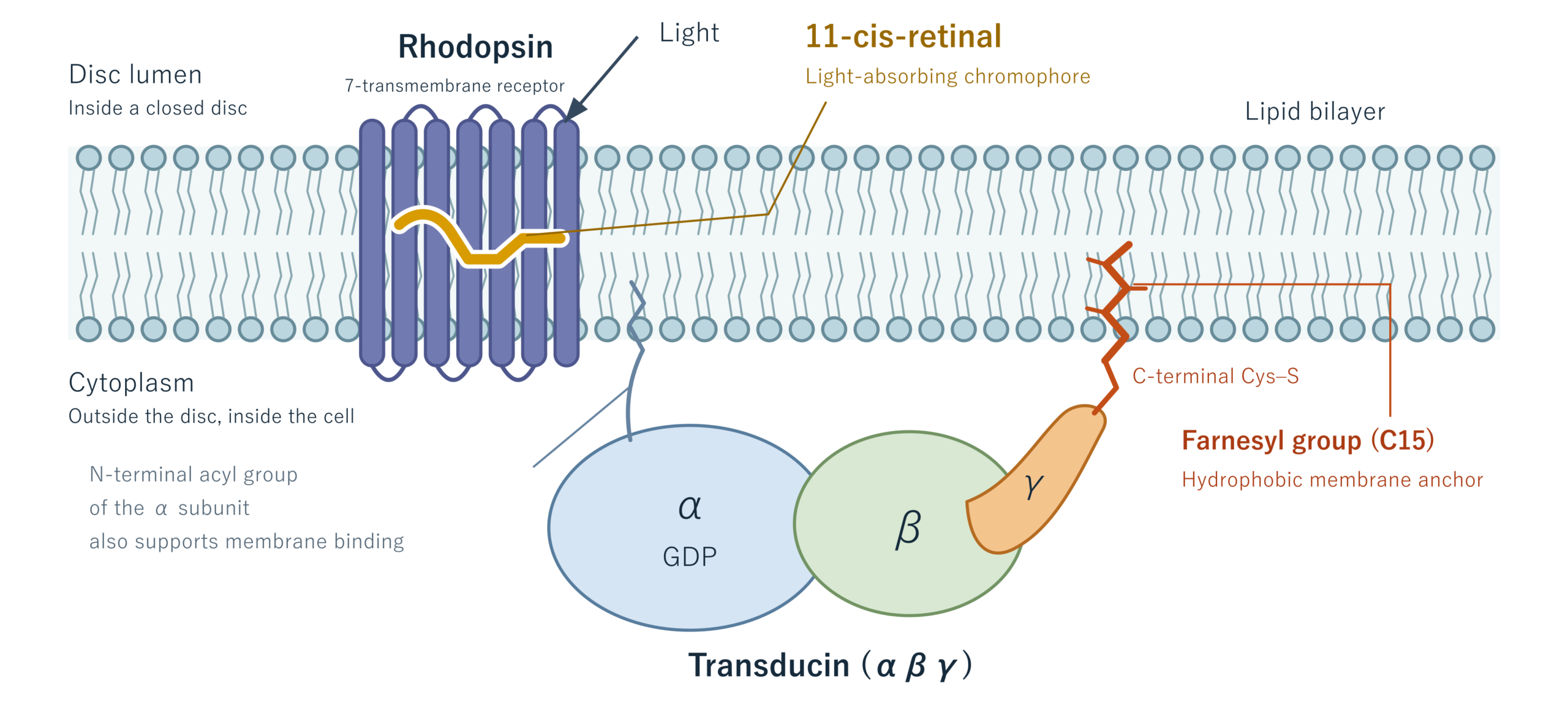
Look at Figure 1. In the outer segment of a rod photoreceptor, flattened, disc-shaped sacs are stacked one on top of another. The space inside a mature disc is the “disc lumen,” and the region outside the disc is the cytoplasm. The lumen is not outside the cell. Rhodopsin spans the disc membrane, while transducin sits on the cytoplasmic side, with the farnesyl group on its γ subunit inserted into the membrane. This “anchor” helps transducin bind to the membrane and receive signals efficiently from rhodopsin. A lipid is also attached to the N-terminus of the α subunit (Tα). However, the lipid modification turned out to differ from the one known in other G proteins. I will discuss this unexpected difference in Part 7.
Having all the necessary proteins in a cell does not, by itself, guarantee that signals will be transmitted successfully. Proteins also need to meet their partners in the right place. The addition of a small lipid helps make these encounters possible.
The Same “Anchor” on a Switch for Cell Proliferation
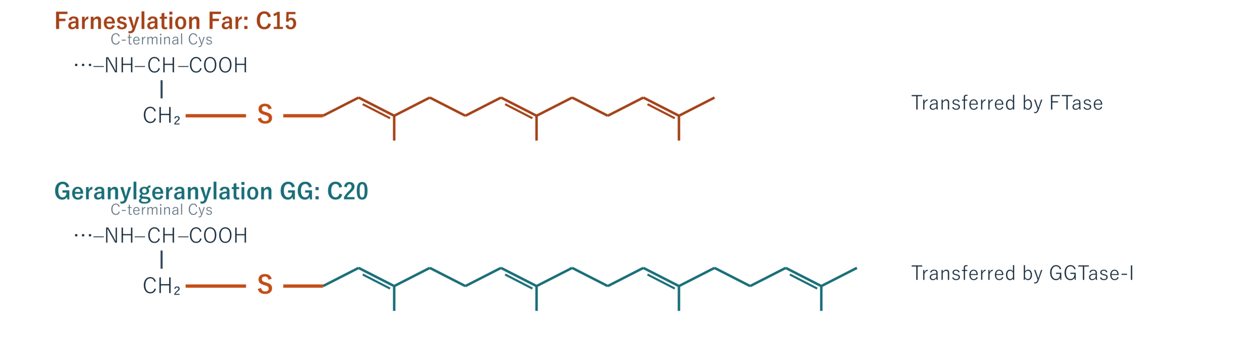
A farnesyl group is a 15-carbon chain. A similar group, the geranylgeranyl group, has 20 carbon atoms (Figure 2). The attachment of these groups is called “prenylation.” Geranylgeranylation is actually more common among the γ subunits of heterotrimeric G proteins. The farnesyl group attached to the γ subunit of rod transducin is the less common form. Enzymes recognize amino acid sequences near the C-terminus of a protein and attach the lipid to the sulfur atom of cysteine (Cys). The resulting C–S–C linkage is called a “thioether bond.”
This mechanism is not limited to vision. Ras, which relays signals for cell proliferation, also carries a farnesyl group. This was reported in 1989, before the report on transducin (2). The lipid modification is also important for Ras to function at the cell membrane.
Members of the Ras family, including K-Ras and N-Ras, act as switches that turn signals for cell proliferation on and off. Certain mutations in their genes can keep these switches on, which can lead to cancer. This prompted research into drugs based on the idea that blocking the attachment of the lipid “anchor” might suppress Ras activity. However, when farnesylation is inhibited, K-Ras and N-Ras can use a geranylgeranyl group instead (3). Even a lipid “anchor” can have a substitute.
Sensing light and promoting cell proliferation are very different processes, yet both involve the same lipid modification. The next time you look at the amino acid sequence of a protein, consider the small lipid that might also be attached to it. In the next article, we will continue exploring how lipids determine where and how proteins work.
1) Fukada Y, Takao T, et al. Nature. 1990;346:658–660. doi:10.1038/346658a0.
2) Casey PJ, et al. Proc Natl Acad Sci USA. 1989;86:8323–8327. doi:10.1073/pnas.86.21.8323.
3) Whyte DB, et al. J Biol Chem. 1997;272:14459–14464. doi:10.1074/jbc.272.22.14459.
Author Profile

Toshifumi Takao
Professor Emeritus, Osaka University; Ph.D. in Science.
From his fourth year as an undergraduate until retirement, he was affiliated with the Institute for Protein Research, Osaka University, where he consistently engaged in research and education in protein chemistry and structural biology.
He currently serves as a Specially Appointed Professor (part-time) at the same institute and as a Life Science Fellow in the Product Division, Rigaku Corporation.
In 2022, he was awarded an honorary doctorate in biochemistry by the University of Havana, Cuba.