Post-translational Modifications Beyond Amino Acids: Glycosylation (Part 4)
Diverse Chemical Modifications Beyond the Genetic Code
In the previous article, we introduced phosphorylation as one of the many chemical modifications that occur in protein molecules. Phosphorylation acts much like a molecular ON-OFF switch. Phosphate groups are repeatedly added and removed within extremely short periods of time, altering protein structures and transmitting biological signals.
In contrast, many of the glycan modifications discussed in this article remain relatively stable once they have been attached. Indeed, many proteins become fully functional only after they have been decorated with glycans, thereby maturing into functional glycoproteins.
Glycans Cannot Be Fully Predicted from Genomic Data
Unlike phosphorylation, the attachment sites of certain glycans can, to some extent, be predicted from genetic sequences. A classic example is N-linked glycosylation at asparagine residues, where glycosyltransferases, the enzymes responsible for attaching glycans, recognize the consensus sequence “Asn-Xaa-Ser/Thr.”
However, the presence of this sequence does not necessarily mean that a glycan will always be attached. As a nascent protein folds into its three-dimensional structure within the endoplasmic reticulum, potential modification sites may become sterically hindered, rendering them inaccessible to the enzyme.
Furthermore, the type and number of attached glycans are often highly heterogeneous, even among molecules of the same protein. Consequently, many questions remain regarding the precise relationship between specific glycan structures and protein function.
Shaping Molecular Space and Modulating Interactions
A defining feature of glycans is the extensive spatial volume they occupy on the protein surface. Compared to linear polypeptide chains, glycans are highly branched and exceptionally flexible. These biophysical properties confer two primary functions.
The first is their role as three-dimensional spacers that maintain appropriate distances both within a protein’s own structure and between the protein and surrounding molecules.
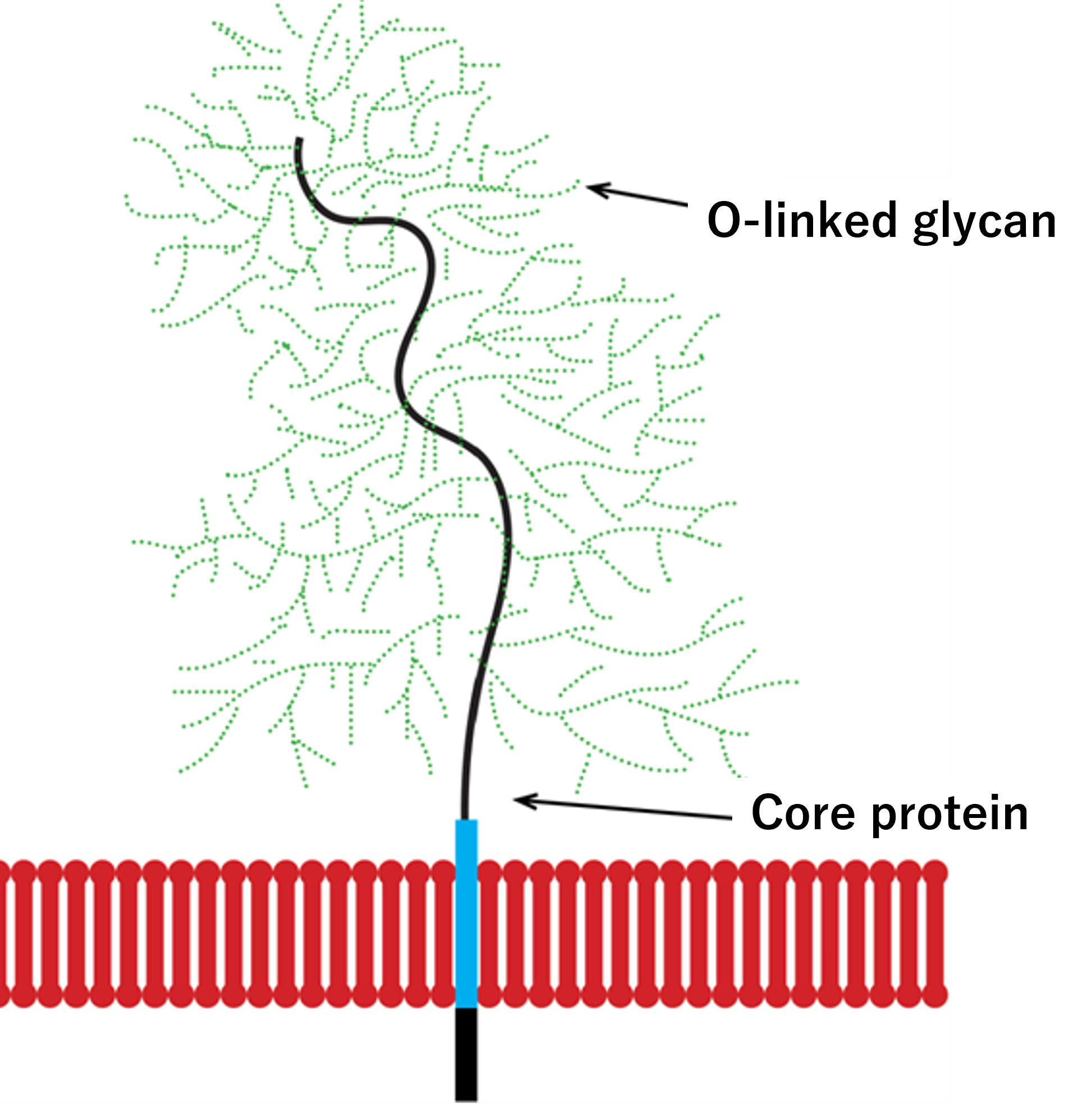
In heavily glycosylated proteins like mucins, dense arrays of glycans surround a central protein core, creating a massive macromolecular assembly. Whereas lipids establish rigid spatial boundaries, as will be discussed in a subsequent article, glycans dynamically fill the surrounding space, engineering a distinct, hydrated molecular microenvironment.
These expansive glycan networks trap large numbers of water molecules and occupy a significant physical volume, effectively functioning as a protective, flexible cushion for cells and tissues. Figure 1 illustrates how the dense glycan shield surrounding the mucin protein core forms this voluminous cushioning layer.
The second major function stems from the unique properties of sialic acid, an acidic sugar frequently capping the outer, non-reducing ends of glycan chains. These terminal structures serve as critical molecular markers, or docking sites, facilitating highly specific biomolecular recognition.
For instance, viral infection by the influenza virus is initiated when its surface protein, hemagglutinin (HA), recognizes and binds to specific sialic acid-containing glycans on the host cell surface.
The crucial determinant of whether an influenza strain preferentially infects birds or humans lies in a subtle structural difference: the linkage of the terminal sialic acid, either an α2,3- or α2,6-linkage. The virus distinguishes these fine structural variations with extraordinary precision.
Avian-to-human transmission is rare primarily because the α2,3-linked glycans preferred by avian influenza viruses are localized deep within the human lower respiratory tract and are sparsely distributed in the nose and throat. This differential anatomical distribution forms a robust “species barrier” to infection.
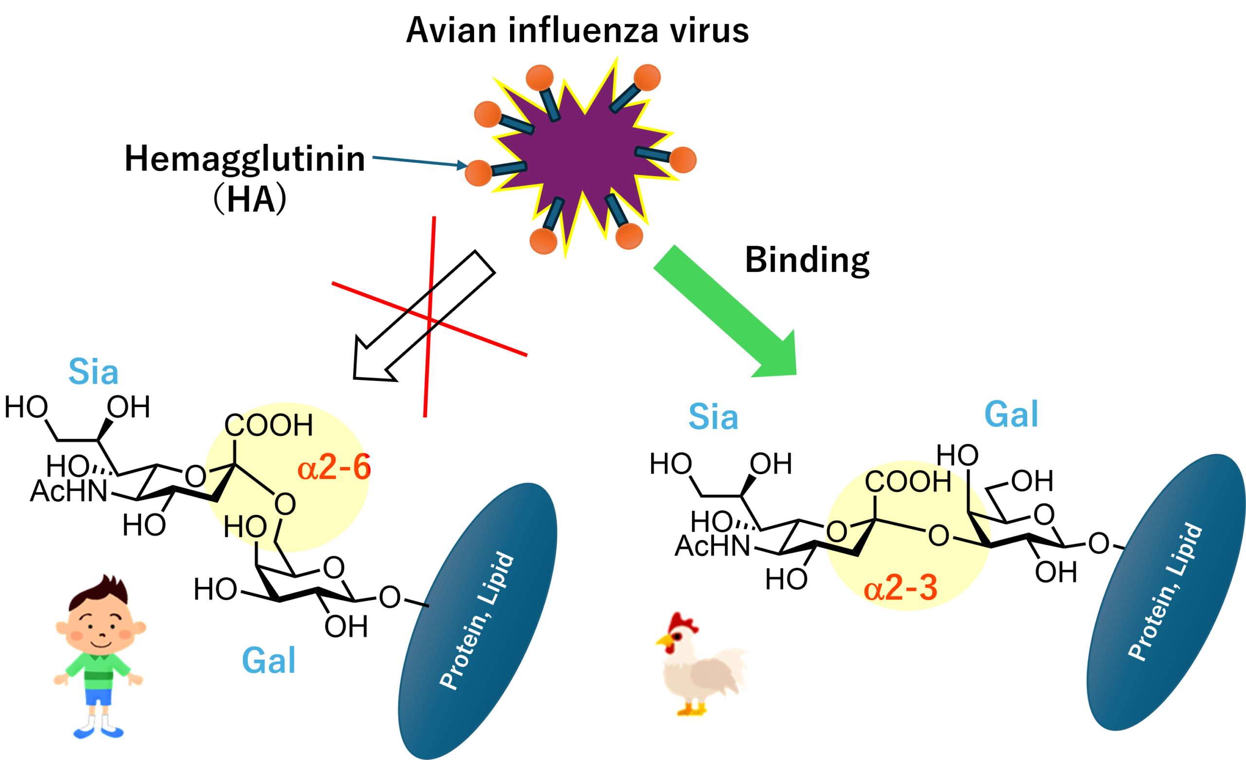
Interestingly, variations in cell-surface glycans also serve as the fundamental basis for the human ABO blood group system.
Tackling 3D Glycan Structures with Advanced AI
These examples represent only a fraction of the diverse roles glycans play in biology. Although their complete structural blueprints cannot be directly read from the genome, their indispensable roles in cellular communication and molecular recognition continue to draw immense scientific interest.
Characterizing the structural diversity and function of glycans remains technically formidable, even with state-of-the-art analytical platforms. Recently, however, advanced artificial intelligence systems, including AlphaFold-driven methodologies, have begun to decode the structural complexities of glycosylation.
Three-dimensional modeling that seamlessly integrates glycans and other post-translational modifications is becoming increasingly feasible. For example, in immunoglobulin G (IgG)—the backbone of modern therapeutic antibodies—conserved glycans attached at specific sites are critical for maintaining the functional conformation of the molecule. Predictive models that fully account for these three-dimensional spatial effects are steadily becoming a reality.
As structural biology and high-resolution analytical technologies continue to evolve, many more of the exquisite structural architectures orchestrated by glycans will undoubtedly come to light. In our next article, we will delve further into the fascinating landscape of post-translational protein modifications.
Author Profile

Toshifumi Takao
Professor Emeritus, Osaka University; Ph.D. in Science.
From his fourth year as an undergraduate until retirement, he was affiliated with the Institute for Protein Research, Osaka University, where he consistently engaged in research and education in protein chemistry and structural biology.
He currently serves as a Specially Appointed Professor (part-time) at the same institute and as a Life Science Fellow in the Product Division, Rigaku Corporation.
In 2022, he was awarded an honorary doctorate in biochemistry by the University of Havana, Cuba.